Friday, November 8, 2013

5 things you must know about Achilles tendon disease

5 things you must know about Achilles tendon disease

Did you know that Achilles tendon ruptures represent a large portion of sports-related injuries and are seen in younger patients and older ones as well.  The average age is 29-40 with a male to female ratio of 20:1. 
In fact, "weekend-warriors" is a term used to classify patients above the age of 35 who participate in occasional exercise activities and especially those that involve high-impact sports such as basketball and soccer.  Those patients are statistically known to be at the highest risk for sustaining Achilles tendon ruptures.
Progression of Achilles Tendon disease

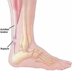
Achilles tendon anatomy.  Image from Gray's Anatomy Text..
Here are Five important facts you must know when dealing with Achilles tendon disease:
  • Achilles tendon disease is progressive - It involves a chronic and repetitive process of micro-tearing and degeneration within the tendon.  This process weakens the tendon and can eventually lead to spontaneous ruptures
  • Pain is not always a clinical symptom of Achilles tendon disease - In fact, the more chronic is the tearing, the less likely pain is involved and thus anti-inflammatory medications would not work
  • Tight calf muscle groups increase the load on the "diseased" tendon
  • Foot-type and mechanics can make you susceptible to Achilles tendon disease
  • Specific family of antibiotics, namely Fluroquinolones, have shown association with Achilles tendon disease and spontaneous ruptures.
Here are important elements to consider for treatment of Achilles tendon disease:
  • Temporary immobilization in a boot/cast is to be expected in the initial "inflammed-state"
  • Shoe-gear modifications, foot-orthoses, physical therapy and stretching exercises are pillars  in the treatment process because they loosen the load off of the tendon
  • Platelet-Rich Plasma injections have shown tremendous promise and results in the treatment especially amongst the athletic/active population
  • Shockwave therapy has also shown great benefits from a non-operative point of view
  • Surgical intervention is warranted if no clinical improvement is seen with non-operative approaches and it involves debridement of the tendon with either a reconstruction of the diseased tendon or a tendon transfer from another tendon in the body. 
    • Recovery involves immobilization in the cast for 3-4 weeks then followed with a period of 4-6 weeks of CAM boot immobilization with Physical therapy
    • Results on average are rated as good to excellent.
If you have any questions about Achilles tendon disease or ruptures, then please consult with your foot and ankle specialist.

Ingrown toenails - 5 important facts and how are they treated?

Have you ever experienced pain, redness, swelling, and even drainage from the "inner" fold of your toenails? Have you been told that this can eventually "grow out"? In reality, ingrown toenails are common in the practice of the foot and ankle specialist but their treatment carries some misconceptions.


Presentation of an ingrown nail with localized infection


Note drainage from inner fold of nail consistent with infected ingrown nails

Here are 5 things you must know about ingrown toenails:
1) Untreated ingrown toenails can cause infections in the skin, bone, and result in life-threatening infections also referred to as "sepsis"
2) Genetics and foot-architecture can play a role for ingrown toenails
3) Cutting toenails too short and into the "corners" can cause ingrown toenails
4) The great (big) toenail is most commonly affected
5) Only definitive way to treat ingrown nails is surgical

Surgical treatment involves an array of different local procedures to remove the "ingrown" component of the nail without compromising the remainder of the nail. In most cases, a chemical treatment is also applied to prevent the nail from growing back "ingrown".

So what does the procedure entail?
1) Typically an in-office setting with local anesthetic infiltrated to "numb" the toe
2) Removing the ingrown component of the nail from the tip to the root
3) Draining and debridement of any infected tissue or abscess.
4) Application of chemical treatment to inhibit nail (matrix) cells from growing back in the in-grown position
5) Application of an antimicrobial dressing that stays on for one day.

Post-operative management?
Although most foot and ankle specialists have different protocols, on average most would agree on a protocol that involves the following:
1) Daily dressing change with a Bandaid until the drainage from the toe completely resolves
2) Following with your provider between 7-14 days after the procedure to ensure no infections. At that point, patients are typically given a prescription for topical solutions to prevent infections and promote the healing of the skin.

If you have any questions about ingrown toenails then please consult with your foot and ankle specialist.

Top 3 reasons why your golf game can be affected by your foot pain

Has your foot pain affected your golf game recently? Have you noticed a change in your swing because of guarding and inability to pivot your foot? The reality of the matter is that there are contributing forces to the golf swing and poor foot mechanics can result in foot pathology that would ultimately affect the quality of the golf swing.


Here are three reasons why your golf swing can be affected by your foot pain:

Great toe joint arthritis - This is a common condition that we see in a large patient spectrum and it is independent of age. The top two reasons that patients develop great toe joint arthritis are: Genetics and/or a history of trauma to the joint. Patients with great toe joint arthritis develop significant pain and stiffness over time, which results in an inability to "push-off" the toe. By guarding the pain, your foot position and mechanics are thereby compromised which leads into a less effective swing.

Plantar fasciitis - This is a condition that involves an inflammatory response at the ligament/tendon structure (plantar fascia), which inserts at the heel. This results in significant pain at the heel and "tightness" in the arch. This, in turn can inhibit the foot from completely planting and thus, will reduce the effectiveness of the swing. We often see this condition in conjunction with tightness of the posterior muscle group which contributes to the pain and inability to plant the heel on the ground.

Achilles Tendonitis - This is a condition that also involves an inflammatory response but this time at the tissues surrounding the Achilles tendon insertion. This is precisely located at the back of the heel and is also seen in conjunction with tightness of the posterior muscle group. The result of this condition is again an inability to plant the foot or push off, which would also compromise the swing.


Here are ways to address those problems:

Stretching
By relieving the contracture and "tightness" of the posterior muscle group
Physical therapy
Can comprise of ultrasound and other hands-on modalities to improve range of motion and relieve tight muscle groups
Mechanical control of the foot - Foot or Ankle and Foot Orthoses
In doing so, there is a lesser tendency or chance of aggravating the tendons.
Vitamin supplementation and antioxidants
Reduce associated swelling and pain
Shock-wave therapy
Using a combination of low intensity ultrasound guided shockwaves, an inflammatory response can be induced in the chronic tendon disease and promote healing.
Surgical repair
Most definitive but is contingent on the type of problem at hand. Typically outpatient type of setting with a brief period of Non-weight bearing (no walking)If you have any questions pertaining to any of the aforementioned conditions, then consult with your foot and ankle specialist

Tuesday, September 3, 2013

What you should know about your feet during pregnancy


Did you know that during pregnancy, your feet experience dynamic changes attributed to fluid retention and increase in soft tissue.  Believe it or not, this is not secondary to stretching, weakness, or ligament laxity.

Here are 5 important facts to know about your feet during pregnancy:
  1. Feet measurements in width and length are increased during pregnancy.
  2. Edema (swelling) in the feet, ankles, and legs are common and can be expected.
  3. Venous insufficiency and varicose veins can be seen in the foot and leg.
  4. Conditioning exercises are not only important to maintain posture but also decrease load on the foot and ankle.
  5. During GAIT, pregnant women have increased demand on the calf-muscle group and over-pronate
In order to help prevent foot problems during your pregnancy, here are three proven methods:
  1. Compression stockings and edema control with elevation
  2. Custom or physician approved orthotics to improve foot function and mechanics
  3. Stretching and conditional exercises to help limit load on the weakened muscle groups
If you have any questions or discomfort with your feet during your pregnancy, then consult with your foot and ankle specialist.

What you should know about ankle arthritis?

Ankle arthritis can be very debilitating and although the prevalence is 9 times lower than knees and hips, the symptoms are much more magnified.  In fact, according to a recent study the quality of life of patients with ankle arthritis was in many cases worse than that of patients with hip arthritis.
Arthritic ankle joint with significant loss of joint space

5 important facts to know about ankle arthritis:
  1. Unlike the hip and knee, ankle arthritis is typically caused by trauma and/or abnormal ankle mechanics
  2. Other less common causes include: inflammatory arthropathies, hemochromatosis, neuropathic arthropathy, bone tumors, and infection.
  3. Cartilage in the ankle joint is much more stiffer and resilient in comparison to the knee joint.
  4. The ankle joint carries up to 5 times the normal body weight.
  5. Both operative and non-operative treatments exist as viable options.
5 important non-operative treatments:
  1. Ankle-Foot-Orthoses (AFO): Function to limit the mobility of the Ankle joint and thereby limiting the associated pain
  2. Corticosteroid injections: Function to limit the associated inflammatory process with motion of the arthritic joint
  3. Physical therapy: Improve GAIT, strengthen muscles, and improve function of contracted tendons secondary to the arthritic joints.
  4. Vitamin supplementation: Antioxidants can help with pain control and associated inflammatory response
  5. Hyaluronate injections: Typically indicated for knee cartilage/joint.  Those injections carry components of healthy joint fluid.  Studies have shown good results in patients with ankle arthritis
Example of an Ankle-Foot-Orthoses  used for patients with ankle arthritis
3 most viable options for operative treatment:
  1. Ankle joint diastasis: Preserves joint and encouraged in early staged arthritis.  Can be useful in select patients.
  2. Ankle joint arthrodesis: Results in complete fusion of the joint, thereby eliminating any joint motion and associated pain.  The body compensates by observing motion at the distal joints of the foot during the GAIT cycle.  It is considered to be the "gold-standard" in surgical management of ankle arthritis since it carries the most predictable of results.
  3. Ankle joint replacement surgery: The joint is completely replaced with a metallic-implant.  It does preserve some limited and pain-free motion of the joint.  It is gaining popularity in the U.S. and is indicated in a very select group of patients.  Newer generation implants carry longer longevity and are more predictable.
Ankle joint replacement with metallic implant
If you have any problems with ankle arthritis, then consult with your foot and ankle specialist.

Wednesday, July 31, 2013

Ankle Sprains

Ankle sprains are a common injury after a fall, sudden twist or blow to the ankle joint. Approximately 40 percent of those who suffer an ankle sprain will experience chronic ankle pain, even after being treated for their initial injury.

A review article published in the May 2009 issue of the Journal of the American Academy of Orthopaedic Surgeons (JAAOS) explains that tendon injuries to the ankle can be a possible cause for this chronic pain. In some cases, the condition is untreated or overlooked which prolongs the pain and the problem. "When patients injure their ankles, the injury may not seem serious at first," explains Terrence Philbin, DO, lead author of the article and Fellowship Director of the Orthopedic Foot and Ankle Center in Columbus, Ohio.

"People may not seek medical attention and they can think it will just get better on its own. I think that is why this condition often goes undiagnosed." The authors of the article describe how in some cases chronic ankle pain may actually be the result of injuries to the peroneal tendons. The peroneal tendons are located behind the outside portion of the anklebone (called the fibula). The tendons help to stabilize the foot and ankle. Tendon injuries can include tendonitis or swelling around the tendons. In more severe cases, the peroneal tendons can actually tear or there can be a swelling of the tendons behind the fibula bone. This can cause the ligament that holds the tendons together to stretch out and tear, or even rupture. Symptoms associated with peroneal tendon injuries can include: Ankle pain that is not responding to treatment Swelling and tenderness around the outside of the ankle Pain behind the anklebone Pain that transmits from the ankle down into the foot.

The use of magnetic resonance imaging (MRI) or ultrasound can be helpful when identifying and diagnosing peroneal tendon injuries and disorders. "These imaging techniques offer a more complete look at the peroneal tendons," noted Philbin. "One might consider getting an MRI or ultrasound especially if you have chronic ankle pain." If the condition is caught early, non-operative treatment options can include: Rest, ice and elevation Anti-inflammatory medication Immobilization in a cast or brace Physical therapy More serious injuries of the peroneal tendons, including tears or ruptures, will very likely require surgery. 

Peroneal tendon injuries can happen suddenly or can develop over time. The injury is most common among athletes involved in sports that require repetitive ankle motion and in individuals who have high arches of the foot. A proper diagnosis is essential in order to treat peroneal tendon injuries correctly and to help alleviate chronic pain. Philbin reminds patients, "If you have ankle pain and it is not getting better, do not ignore it. Get it evaluated by a physician who has experience treating foot and ankle injuries."

We can try a variety of measures to reduce pain and swelling and disability from this condition. PRP injections, low profile bracing, and physical therapy are excellent nonsurgical options. Ultimately surgery would be considered pending the nature and severity of the problem, and how you respond or fail to respond with nonsurgical management. 
New data gleaned from five years of studies have shown that artificial sweeteners can lead to weight gain as the body forgets how to process real sugar.


Sweet taste without the guilt — this has long been the promise of diet sodas.

But researchers at Purdue University have found by reviewing studies done over the past five years that diet soda and other artificially sweetened products could cause the same health problems as regular soda. In review, Susan E. Swithers, a Purdue professor of behavioral neuroscience, discovered that with consuming artificial sweeteners like sucralose, aspartame and saccharin, the body loses its ability to process real sugar, which could result in weight gain.

With real sugar, messages are released to the brain and the gullet via hormones that regulate digestion and intake of food, blood sugar levels, and even perception of fullness and satisfaction from the food consumed. However, with no actual calories to process with artificial sweeteners, the body doesn't know how to react.
"You've messed up the whole system, so when you consume real sugar, your body doesn't know if it should try to process it because it's been tricked by the fake sugar so many times," says Swithers to CNN.

The end result, says Swithers, is that the body's metabolic system stops reacting to real sugars and doesn’t release the hormones that say, “That was delicious, I’m full.” This can cause an endless cycle of feeling hungry and wanting more sugar.  It has also been shown that people who regularly drink diet soda have a higher risk of weight gain, obesity and type 2 diabetes and heart disease.

However, representatives of the American Beverage Association disagree and argue that low-calorie products can be effective tools for weight control, calling the study “an opinion piece,” in a released statement, according to CNN.

Theresa Hedrick, nutrition and scientific affairs specialist for the Calorie Control Council lobbying group, told WebMD, "I think it's important to remember that low-calorie sweeteners are one aspect of a multifaceted approach to health or obesity prevention. ...They aren't magic bullets."

Weight gain is a major contributor to all foot and ankle problems, and our patients need to understand that weight loss therefore will directly impact the reduction of most foot and ankle pain. Ask us about your weight loss options today, or at your next appointment.

Friday, July 19, 2013

What you should know about Gout in the foot and ankle


Have you ever suffered from a painful and swollen joint in your foot or ankle?  Has this pain persisted despite taking antibiotics and anti-inflammatory medications?  Have you had multiple episodes of this pain?  If so, then you may be one of 8.5 million Americans who suffer from gout each year (1).

Prevalence of Gout has been increasing in recent years and it is currently one of the most common causes of inflammatory arthritis in industrialized countries.  Factors that contribute to this condition include increased lifespan, dietary habits, renal and cardiovascular disease, alcohol consumption, and prescribed drugs that may raise serum uric acid levels (2).

Gout results from the deposition of uric acid crystals in a joint, resulting in acute inflammatory response.  If left untreated, chronic gouty arthropathy can develop and progress into deposition of uric acid crystals and tophi in soft tissues.  In the foot and ankle, Gout is most commonly manifested in the great toe joint.

Current treatment guidelines:
  • Non-Steroidal Anti-inflammatory medications (NSAIDS) or corticosteriods in the acute phase
  • Uric acid Lowering Treatments (ULT), i.e. Allopurinol, for maintenance
  • Shoe gear modification – wider, supportive shoes with insoles
  • Custom Orthotics – accommodations to restrict motion of the affected joints
  • Ankle Foot Orthoses (AFO) – restrict motion of the ankle joint (if affected) to limit inflammatory response
  • Vitamin supplementation
  • Surgical reconstruction – reserved for failed conservative and pharmaceutical therapies

If you suffer from gout or symptoms resembling gout, then consult with your foot and ankle specialist for work-up and management.

Foot sprains - Are they more than just sprains?


Have you ever been diagnosed by your doctor with a “foot sprain” after an accident, fall, or foot-twist?  Were you immobilized with a boot for a short period of time without much relief?  The reality of the situation is that you may have incurred a more debilitating injury in your foot ligaments. 

Some might ask what do the National Football League athletes Santonio Holmes, Ryan Kalil, Matt Schaub, and Dwight Freeney have in common?  They all suffered from a frustrating foot injury commonly referred to as Lisfranc’s injury.

The injury is named after Jacques Lisfranc, a French surgeon in Napoleon’s army.  It refers to a ligament and/or osseous (bone) injury of the metatarsal-tarsal joints and can present with severe dislocations and fractures (1).  The mechanism of the injury involves either a direct force of trauma on a planted foot or indirectly through a complex sequence of twisting motion.

Diastasis noted in the joint complex consistent with Lisfranc ligament injury
Approximately 20% of Lisfranc injuries are misdiagnosed in the emergency room because of their vague presentation and subtle x-ray findings (2).  They are often mislabeled as "foot sprains” and thus, treatment is often delayed which results in:
  • Post-traumatic arthritis
  • Chronic midfoot instability
  • Chronic pain
Lisfranc injuries occur in 1 person per 55,000 yearly.  67% of which are results of motor vehicle accidents, crushing injuries, and falls from heights (2).  Although those injuries are not common in the general population, certain athletes suffer a higher rate of this injury.  It is seen in 4% of football players per year, with offensive linemen incurring 29.2% (3). 

Foot and ankle specialists are trained to recognize the pathology using specific clinical examination and with the help of imaging modalities such as stress x-rays, CT scans, and even MR images.  If recognized early, prompt treatment can limit complications and potentially salvage an athlete’s season/career.

If you have sustained or suspect a foot sprain, then consult with your foot and ankle specialist.  

Monday, June 3, 2013


Unseen Dangers of Walking Barefoot on the Beach


Do you enjoy long, barefoot walks on the beach? While it may be romantic, it can be potentially dangerous. Shell fragments or other sharp debris can penetrate your skin causing cuts or puncture wounds that can be extremely painful. If not treated properly, they can become infected by a number of bacteria.

One specific bacterium of concern is Vibrio vulnificus. Infections related to this bacterium typically happen when a cut or wound occurs in salt or brackish water. This organism peaks during the warm summer months when the gulf coast water surface temperature exceeds 68° F. High concentrations of Vibrio are commonly found in filter-feeding organisms such as barnacles, and therefore any cuts or scrapes from barnacles are at an increased risk for infection. Other possible sources of infection include punctures from fishhooks and fish spines or teeth. Following exposure to Vibrio, a local skin infection with large blisters may occur. In an individual with an underlying illness such as liver disease or diabetes, the infection may spread extremely rapidly resulting in significant tissue damage, and if Vibrio infects the bloodstream, it is potentially fatal. Treatment for infection includes antibiotics and possible hospitalization and surgical debridement depending on the severity of the wound.

When it comes to cuts or wounds involving salt or brackish water, it is best to be safe and seek medical attention.
Skin Cancer and your feet..

Most people are unaware of every little pigmented lesion they may have on their foot or toes. Sometimes, a pigmented lesion may be falsely presumed to be a bruise, or a scar. If there are any questions about skin lesions, one should note that a skin biopsy is a relatively small amount of discomfort to gain a relatively large amount of information and essentially a diagnosis which can be used to effectively treat any number of skin cancers which may present as old bruises underneath a thick toenail, or even a rash on the top of the foot. Please understand, that although relatively rare (5% of all skin cancers such as melanoma are found on the foot) this is still high enough to get it looked at.

Monday, May 6, 2013


Overview of Peroneal Tendon Issues


Many patients that we treat at our office have chronic ankle instability in the form of lateral tendon dislcocations, or chronic ankle sprains. Many times these patients are unclear as to how important these tendons are to the overal stability and function of the ankle joint. With abnormal tendon gliding and ligamentous attenuations and ruptures, these tendons may also become painful with patients who have chronic ankle sprains. This is a comprehensive overview of this pathology and treatment options to help out with the understanding of these clinical scenarios.

History of the Procedure
Disorders of the peroneal tendons have been reported infrequently. Monteggia described peroneal tendon subluxation in 1803, and this entity seems to be more commonly encountered than are disruptions of the peroneus longus or brevis alone. Nonetheless, peroneus brevis disorders have been described more often in the literature, with peroneus longus problems gaining more recent attention. However, much of the literature regarding both tendons is in the form of case reports.

Problem
The peroneal muscles make up the lateral compartment of the leg and receive innervation from the superficial peroneal nerve. The peroneus longus muscle originates from the lateral condyle of the tibia and the head of the fibula. The tendon of peroneus longus courses behind the peroneus brevis tendon at the level of the ankle joint, travels inferior to the peroneal tubercle, and turns sharply in a medial direction at the cuboid bone. The tendon inserts into the lateral aspect of the plantar first metatarsal and medial cuneiform.
A sesamoid bone called the os peroneum may be present within the peroneus longus tendon at about the level of the calcaneocuboid joint. The frequency with which an os peroneum occurs is controversial, with many supporting the idea that one is always present. However, the os peroneum may be ossified in only 20% of the population. The peroneus longus serves to plantar flex the first ray, evert the foot, and plantar flex the ankle.

The peroneus brevis originates from the fibula in the middle third of the leg. Its tendon courses anterior to the peroneus longus tendon at the ankle. It courses over the peroneal tubercle and inserts onto the base of the fifth metatarsal. The peroneus brevis everts and plantar flexes the foot.

Problems may arise in either of the tendons alone, or both may be involved with subluxation. The hallmark of disorders of the peroneal tendons is laterally based ankle or foot pain. Whether the problem is tendinous degeneration or subluxation, the clinical manifestation is pain. With time, loss of eversion strength may occur.

Problems arising with the peroneus longus include tenosynovitis and tendinous disruption (acute or chronic). The os peroneum may be involved with the degenerative process or as a singular disorder and can be fractured or fragmented. Longitudinal tears of the peroneus longus are uncommon but have been reported.
Longitudinal tears of the tendon are the most common problem seen with the peroneus brevis tendon. These may be single or multiple. Tendinitis and tenosynovitis also may occur.

Subluxation of both peroneal tendons may occur following an acute traumatic episode or may be of a more chronic nature.

Frequency
Disorders of the peroneal tendons are less common than other tendon problems involving the Achilles or posterior tibial tendons. However, it is impossible to estimate their true frequency in the United States or abroad.

Etiology
The precise etiology of peroneal tendon disorders depends somewhat on the specific problem being addressed. All disorders may result following a traumatic episode, direct or indirect, with a lateral ankle sprain being the most common trauma. Brandes and Smith have reported that 82% of patients with primary peroneus longus tendinopathy had a cavo-varus hindfoot.3 The presence of an os peroneum also has been postulated to predispose to peroneus longus rupture. Ruptures likewise have been reported to occur secondary to rheumatoid arthritis and psoriasis, as well as diabetic neuropathy, hyperparathyroidism, and local steroid injection.4,5,6
Longitudinal splits in the peroneus brevis tendon appear to result from mechanical factors. Repetitive or acute trauma causes the attritional ruptures. These ruptures may result from an incompetent superior peroneal retinaculum that allows the peroneus brevis to rub abnormally against the fibula.

Overcrowding from a peroneus quartus muscle also has been reported. The blood supply to the tendon has been shown to be adequate.

Subluxation of the peroneal tendons results from disruption of the superior peroneal retinaculum and usually involves avulsion of the retinaculum from its fibular insertion. The mechanism of injury typically involves an inversion injury to the dorsiflexed ankle with concomitant forceful contraction of the peroneals. Some patients have a more chronic presentation and cannot recall a traumatic episode. Congenital dislocations also have been reported. An inadequate groove for the peroneals in the posterolateral fibula may be a cause of subluxation as well.

Pathology of the longus and brevis tendons almost always occurs concurrently. Brandes and Smith noted a 33% incidence of concomitant problems.

Presentation
The patient with peroneal tendon pathology typically complains of laterally based ankle or hindfoot pain. The pain usually worsens with activity. However, presentation and diagnosis often are delayed. Patients may or may not recall a specific episode of trauma. Brandes and Smith reported that only 9 of 22 patients with primary peroneus longus tendinopathy recalled an inciting event and that the event was an average of 4.3 months prior to presentation.

Peroneal tendon subluxation or dislocation may present acutely following a traumatic injury to the ankle. However, it is not uncommon for these to present later with an uncertain history of trauma. Patients also may complain of snapping or popping in the ankle.

On physical examination, there usually is tenderness to palpation along the course of the peroneal tendons. Edema also may be present. These disorders require a high level of suspicion. Even frank dislocations may be missed if not specifically evaluated.

A provocative test for peroneal pathology has been described. The patient's foot is examined hanging in a relaxed position with the knee flexed 90º. Slight pressure is applied to the peroneal tendons posterior to the fibula. The patient is then asked to forcibly dorsiflex and evert the foot. Pain may be elicited, or the tendons may be felt to sublux.

Indications
The primary indication for treating these disorders is pain. Nonsurgical treatment usually is attempted first. Failure of conservative measures is an indication for operative intervention.

Operative Considerations

With physical therapy, MRI, and need for primary or secondary repair will be determined based on overal health of the patient, as well as how effective nonsurgical measures have been. If the pain and resolution is not fully noted through physical therapy and bracing one should consider the possiblity of repair. If there are tendon tears associated with the pathology, repair is recommended. If an associated low muscle (peroneus quartius) or ruptured retinaculum is identified, repair is also likely required. We are experts in this pathology, and treat this regularly, and I feel a proper evaluation for this condition will be beneficial to anyone with recurrent ankle sprains, as well as pain in the lateral ankle.


Hammer Toes


Hammertoes occur when the smaller toes of the foot become bent and prominent. The four smaller toes of the foot are much like the same fingers in the hand. Each has three bones (phalanges) which have joints between them (interphalangeal joints). The toes form a joint with the long bones of the foot (metatarsals) and it is this area that is often referred to as the ball of the foot.
Normally, these bones and joints are straight. A hammertoe occurs when the toes become bent at the first interphalangeal joint, making the toe prominent. This can affect any number of the lesser toes. In some cases, a bursa (rather like a deep blister) is formed over the joint and this can become inflamed (bursitis). With time, hard skin (callous) or corns (condensed areas of callous) can form over the joints or at the tip of the toe.


What causes hammertoes?
There are many different causes but commonly it is due to shoes or the way in which the foot works (functions) during walking. If the foot is too mobile and / or the tendons that control toe movement are over active, this causes increased pull on the toes which may result in deformity.
In some instances trauma (either direct injury or overuse from walking or sport) can predispose to hammertoes. Patients who have other conditions such as diabetes, rheumatoid arthritis and neuromuscular conditions are more likely to develop hammertoes.

Are women more likely to get the problem?
It is more common in women as they tend to wear tighter, narrower shoes with increased heel height. These shoes place a lot of pressure onto the joint and predispose to deformity. It is common for patients to wear shoes that are too small and this can predispose to the problem. In a study we have performed, 95% of patients were in the wrong size shoes.

Will it get worse?
At the start of the deformity, it is generally mobile which means that the toe can be straightened. However, with time, the joint become fixed or rigid. This can then affect the joint at the ball of the foot and, in severe cases, the joint capsule ruptures (tears) so that the joint becomes dislocated and the toe sits up in the air.

What are the common symptoms?
Deformity / prominence of toe
Pain
Redness around the joints
Swelling around the joints
Corn / Callous
Difficulty in shoes with deformity of the shoe upper
Difficulty in walking
Stiffness in the joints of the toe

How is it identified?

Clinical examination and a detailed history allow diagnosis. X-rays are often not required but can help to evaluate the extent of the deformity and the degree of arthritis within the joint.

What can I do to reduce the pain?
There are several things that you can do to try and relieve your symptoms:
Wear good fitting shoes with a deep toe box
Avoid high heels
Use a toe prop to straighten the toe if it is still mobile
Wear a protective pad over the toe
See a doctor at the Family Foot and Leg Center.

What can we as a specialist do to correct or reduce your symptoms?
If simple measures do not reduce your symptoms, there are other options:
Advise appropriate shoes
Advise exercises if the toes are still mobile
Show you how to strap the toe in a corrected position
Provide a splint or protection
Consider orthotics

Advise on surgery
The way in which your foot loads during walking can place increased stress on the ball of the foot and cause increased toe activity. Special shoe inserts (orthoses) can help to control foot movement. Whilst these are unlikely to resolve established deformity they may help reduce discomfort in the ball of the foot.

Will this cure the problem?
If the deformity is mobile, then this may help prevent progression although there have been no scientific studies to analyse the benefit. If the deformity is fixed, then orthotics will not cure the problem but may reduce the associated symptoms.

What will happen if I leave this alone?
Generally, the deformity becomes worse with time and slowly becomes fixed (stiff). This can cause discomfort in shoes. The position of the toe places increased stress on the ball of the foot and this can become painful. Corn and callous formation on the ball of the foot is not uncommon. In some cases, the metatarsophalangeal joint capsule ruptures, causing the toe to sit up in the air.

Can the deformity be reversed or cured?
The only effective way of correcting the deformity is to have an operation.

How does the operation correct the deformity?
There are a number of different operations. However, the most common operations are:
Tendon transfer
Digital arthroplasty
Digital arthrodesis


Hopefully this is an effective run down of various questions commonly asked by my patients here, and if you come up with more please comment and the questions will be answered.

Tuesday, April 23, 2013


Baby and Toddler Shoes


Before babies starts walking, they don't need shoes. In fact, supportive shoes like hard-soled Mary Janes may actually get in the way of your child's developing mobility. Socks, booties, and soft-soled baby shoes are useful for warmth, but bare feet are fine, too. In actuality their feet are very adaptable to the ground, whether it is carpeted or wood and tile. And early on it allows their proprioceptive feedback to allow them to balance easier and develop more readily in the very early stages without shoes. 

Once your child takes those first steps, it's time for a pair of real shoes. Unlike "baby shoes," which are more like slippers, first shoes will have a flexible, nonskid sole (probably rubber) and a more substantial upper. Shoes protect kids' feet outdoors and anywhere else that could be hazardous – a splintery surface, for example.

Note: Your child's foot is still developing, so it won't look (or act) like an adult foot. If your child still has a padding of baby fat under the arches, for example, she might appear a bit flat-footed. Or she may have a tendency to turn her toes in when she walks, called in-toeing or toeing in.Indoors (and outdoors on safe surfaces, such as sand), it's still a good idea to let new walkers wear soft baby shoes or socks. Your child can even go barefoot, if it's warm enough. Toddling around with feet bare or lightly covered actually helps little ones build strength and coordination in their legs and feet.
Mention any concerns to your child's doctor. It's easier to correct foot problems when your child is younger.


What to look for when buying
Choose a breathable, lightweight material. Soft leather or cloth is best. Avoid stiff leather shoes, which can hinder foot development, and synthetics, which don't breathe. Bend the soles. They should be flexible and gripping, not smooth and stiff. A nonskid rubber sole with ridges will offer good traction.

Check the fit. Have your child try on the shoes and stand up. There should be just enough room to squeeze your pinky between your child's heel and the heel of the shoe, and a full thumb-width between the end of your child's longest toe and the front of the shoe. The shoe should provide just enough wiggle room without being too big. Because baby feet grow quickly, it's a good idea to check every month to make sure the shoes still fit.
Give it a squeeze. If the shoe is made of soft fabric, try to grab some of the material on the top of the foot when your child is wearing them. If you can't, the shoes might be too tight.
Shop later in the day. Babies' feet swell and are often bigger at the end of the day. Shoes purchased in the morning might feel tight in the evening so usually try shoes on before dinner before you buy.
Look for problem spots. Your baby's shoes shouldn't need any breaking in. Let your child toddle around indoors wearing the shoes, then take them off and look for any irritated areas on your child's foot which could be red areas or blisters, and in more rare cases even a callus formation can occur but this is not common.
Make the choice: laces versus Velcro. Velcro fasteners make it easier to get shoes on and off, and you won't have to worry about retying laces all day. But a child may figure out how to remove his shoes and take them off when you wish he wouldn't! If you choose shoes with laces, make sure they're long enough to tie into double knots, so they won't come undone as often.


Achilles Tendonitis

The Achilles tendon is the thickest and strongest tendon in the human body. This tendon, which runs along the back of the leg and inserts onto the heel, can endure forces of up to 6 to 8 times the body weight during repetitive activities such as running.  Because of the high load of stress it must endure, the Achilles tendon is prone to injury and inflammation. This condition is known as Achilles tendinitis and is a common cause of leg and heel pain in the active individual. 

There are many factors which may contribute to the development of Achilles tendinitis, but it is commonly an overuse injury which results from a sudden change in activity level without proper training or conditioning. Other factors include tight leg muscles, improper shoes, and biomechanical faults within the foot and leg. 

Signs of Achilles tendinitis often begin with swelling and a dull ache or stiffness in the back of the leg and heel that typically occurs at the end of activities. If left untreated, the pain can worsen and become present at the start of activities or even during normal walking. In severe cases, the tendon may even partially or completely rupture. 

Early cases of Achilles tendinitis can be treated conservatively with rest, ice, and gentle stretching. Orthotics and heel lifts may help relieve tension on the tendon. Physical therapy can be initiated to provide additional reduction of inflammation and pain. In some cases, the Achilles tendon may require temporary immobilized within a walking boot. 

Surgery may be necessary for individuals with pain that persists or worsens despite conservative treatments. However, it may be possible to avoid surgery through the use of advanced therapies such as extracorporeal shock wave therapy or platelet gel injections. Once the pain resolves, it is important to have a gradual return to activities to avoid re-aggravating the Achilles tendon. 

Participation in a regular physical activity is an important part of maintaining a healthy lifestyle. Proper shoes, stretching, and strengthening exercises can help prevent injuries. For any foot or ankle pain, a podiatrist should be seen for a full assessment and treatment.

Sunday, April 14, 2013

Treatment and Prevention of Blisters


A common complaint athletes have is the formation of blisters on their feet. They arise in areas that are subjected to excessive and repetitive friction. In addition, heat and moisture contribute to blister formation by softening the outer layer of skin. These factors are all present during activities such as running. While blisters typically are a painful nuisance, they may develop into an infection if not properly cared for.

Some blisters will resolve if left alone, but if the blister is painful, it may be drained. Using rubbing alcohol, sterilize a needle and the skin over the blister. Carefully lance the thin, outer layer of the blister and drain the fluid. Apply an antiseptic (iodine or antibiotic ointment) and cover with a bandage. Continue to watch for any signs of infection over the next few days as the blister heals.

If you develop recurrent blisters, you may treat them after they form; however, a better solution is to prevent their formation.

There are numerous products available that can decrease the coefficient of friction. These products (Body Glide, Bag Balm, or petroleum jelly) act as a lubricant when applied to the skin.

If your feet sweat a lot, your skin will soften over time increasing the likelihood of a blister. Foot powders can help absorb excess moisture keeping your skin dry and intact. Spraying your feet with an antiperspirant is another easy option.

The next step in prevention is wearing proper socks which should be made from a synthetic, moisture-wicking fabric. Consider wearing a double-layered sock (WrightSock) as it reduces friction against your skin. If you tend to develop blisters between your toes, a toesock (Injinji) provide extra protection.

And finally, the most common cause of blister formation is improperly fitting shoes. Make sure your shoes fit properly around your heel and that there is plenty of room in the toebox. Shoes that are too long or wide can cause your heel to slide and lead to a heel blister. Shoes that are too short or narrow increase the frictional forces upon your toes.

The best treatment of blisters is pro-active prevention. By following these suggestions, blisters do not have to be a part of your sport or exercise routine.

Time for New Shoes?

You’ve finally found the perfect pair of running shoes. They fit and feel wonderful. You feel as if you can run forever in them, but they are now starting to show some wear. Is it time toss your favorite shoes and get a new pair?


There are several things to consider when deciding if your athletic shoes need to be replaced. First, how much has the shoe been used? For high-impact activities such as basketball or tennis, shoes should be replaced after roughly 60 hours of use. A simple guideline for running shoes is to change them every 300-500 miles, but there are many factors than can shorten or extend the life of a shoe. For example, a petite runner will have less wear than a heavy runner. The type of running surface can also make a difference as a soft trail will result in less wear than a concrete sidewalk.


Next, check for signs of wear on the outsole. Place your shoes on a flat surface and examine them from behind. Excessive wear may cause the shoe to tilt to one side. If the heels are not perpendicular to the ground, the shoes need to be replaced. For most heel-striking runners, a normal wear pattern is on the outside corner of the heel. If one typically runs on a road or track with a cant (cross slope), an asymmetric wear pattern may develop. A significant asymmetry should be evaluated by a podiatrist as it may be caused by an issue that may be improved through the use of a custom orthotic. 

Finally, it is important to know that the cushioning and support of a shoe will eventually wear down, even if the visible exterior remains in good condition. Athletic shoe midsoles are typically made of EVA which is a compressible material that loses its shock absorptive capabilities with prolonged use. Other factors can speed the degradation of EVA such as high heat or UV light. Loss of cushioning can lead to injuries such as stress fractures or shin splints. Another part of the shoe that can wear out is the upper which holds the shoe onto the foot. This supportive portion of the shoe can become over-stretched with use. When this occurs, the foot has more room to move inside the shoe which can lead to injuries and sprains. 

Good shoes are important in all activities as they provide support, stability, and shock absorption. With continued use, the loss of these protective functions can potentially lead to overuse injuries of the foot and leg such as plantar fasciitis, stress fractures, ankle sprains, and shin splints. The decision to replace your shoes depends on several factors, but if your shoes appear worn, it is time for new shoes.

Tuesday, March 26, 2013

Runner's Knee


A new course record was set at the Chicago Marathon last year.  With a time of 2:04:38, Tsegaye Kebede became the first Ethiopian man to win on Chicago’s flat course.  Thousands of first-timers and other runners followed the elite runners this morning.  For these endurance athletes, training is essential to reach the finish line, but overtraining or overpacing can easily lead to injury.  One common running injury is known as runner’s knee (patellofemoral pain syndrome) which is associated with pain behind the kneecap.  It is an overuse injury often seen in runners and cyclists, but it can also be seen in sports which require repetitive jumping or cutting.  While this pain can be cause by a direct impact injury, it is typically caused by repetitive bending of the knee.
 
The major symptom is pain which usually begins as a dull ache or stiffness behind the kneecap (patella).  The patella is attached to the thigh by the quadriceps muscle and tendon and to the leg by the patellar tendon.  Injuries or weakness of these supporting structures can lead to improper alignment and tracking of the patella as the knee  bends and straightens.  This results in irritation and pain.
 
Conservative treatments for patellofemoral pain syndrome begin with reduced training, ice, and anti-inflammatory medication.  Incorporate stretching and strength training of the quadriceps and hamstrings to improve stability around the knee.  In addition to these therapies, custom arch supports (orthotics) may be considered.
 
Recent studies have provided evidence that orthotics reduce patellofemoral pain by improving the tracking of the patella during the bending motion of the knee.  Although orthotics have been generally thought to benefit individuals with excessive collapsing of the arch (over pronation), these studies have shown that people of all foot types have reduction in knee pain with orthotics when compared to flat inserts.
 
Patellofemoral pain syndrome is a degenerative condition that can progressively get worse over time.  Proper training and conditioning can help prevent this and other lower extremity injuries.  Following any injury, the goal of returning to activity as quickly and safely as possible can be achieved with proper evaluation and treatment.  Custom orthotics casted by a podiatrist can be an important addition to the standard treatments for runner's knee.

Monday, March 25, 2013

Big Toe Joint Aches and Pains - What you need to know:


Often times patients will present with what they consider to be a "Bunion" in their big toe joint, and they attribute this to their pain. Many times, this is false. A bunion is an actual deviation or dislocation of the big toe joint, whereas the painful prominence which are often gradually increasing in size after many years removed from an injury can be related or directly attributed to progressive "bump" production which has the general buzzword "Arthritis".

Arthritis is actually a misnomer, because many times the inflammation of the joint is only noted initially in the disease process and over time the cartilage wears away forming spurs and bone prominence which leads to the pain and inability to move the joint fluidly. This is actually an "arthrosis" which is the end result of the condition. Arthritis is not usually visualized directly on a radiograph but arthrosis is easily visualized.

A clinician will find effusions whenever arthritis is active. This is the "Swelling" that patients often note is painful and warrants medical attention. This is often including joint space widening in the case of inflammatory arthritis but narrowing over time with osteoarthritis, and reduction of ability to move the joint without pain and with eventual grinding.

Patients will often times not recall the traumatic event which sparks the progressive wear down of this area on their foot, but many times this is not necessary. Patients will also note that the "bunion" is growing and is usually located on top and side of the joint. This is not a bunion, as these tend to only be located on the side of the joint and not at all on top.

What do you need to know to determine what to do next??

1) If the pain is at the end of the day you should see a foot and ankle specialist.

2) If the motion of the joint is limited or not similar to your other foot, you need to see the specialist.

3) If you have been given 2 or more cortisone injections in the same joint, you need to see a specialist.


There is a common theme here. A foot and ankle specialist is paramount to help a patient achieve a good result in treating first toe joint arthrosis. Often times, this is progressive and may limit treatment options depending on the stage of the pathology. There is no actual cure for any form of arthritis, but there are means to treat and accommodate the condition in order to reduce pain and limitation with walking. We are adept in treating all forms of arthritis, from the initial swelling stage, to the end stage limited motion in the toe joint. There are many times several good options depending on the stage of the disease process, and we will be able to give you a definitive solution in many cases.

Advancement in Achilles Tendon Tear and Achilles Rupture Care

The Achilles tendon is the strongest tendon in the body but it is also the one that is most commonly injured. In this article we discuss the anatomy of the Achilles tendon as well as give an overview on Achilles tendon ruptures.
 
Achilles Tendon Anatomy

The Achilles tendon is approximately 15 cm in length and is made up of a combination of fibers that are derived from two calf muscles which twist 90o and insert at the back of the calcaneus or heel bone. The Achilles tendon gets its blood supply from 3 sources. There is an area on the Achilles tendon approximately 2 to 6 cm above its insertion to the calcaneus that has poor circulation and is called the watershed region. Due to the diminished blood supply, this is a common area for Achilles tendon lesions and ruptures.


Acute Achilles tendon ruptures

Achilles tendon ruptures occur most often in middle-aged men during athletic activities. Patients with Achilles tendon ruptures often say that they felt a sudden pop or snap in their calf with subsequent weakness or difficulty with ambulation. Some say they felt someone kicked them or hit them from behind but there is noone in the vicinity. Patients may be able to walk with minor swelling and pain after a tear but have weakness.
Achilles Tendon Tear Exam

On examination, a palpable defect in the tendon can be felt and there is often an increase in upward movement of the ankle due to the lack of tension from the Achilles tendon. A common test that is used to evaluate the integrity of the Achilles tendon is the Thompson calf-squeeze test. Squeezing the calf muscles on the affected side will yield little to no movement in the ankle when compared to the unaffected side.
Imaging of Achilles Tendon Tears or Ruptures

MRI and ultrasound are often used to confirm the presence of an Achilles rupture and to evaluate the extent of the rupture. The diagnosis of Achilles tendon ruptures, however, is based on clinical examination. Imaging is often used for surgical planning or in cases in which there are equivocal examination findings. 
 
Achilles Tendon Tear Surgical Advancements

Operative and nonoperative methods offered by foot and ankle doctors have shown favorable outcomes. Generally with surgical repair, there have been reports that there is a decreased rate of re-rupturing the tendon, improved strength, improved ankle motion, better return to activities, and fewer complaints.

Operative treatment involves reapproximating the two ends of the ruptured tendon together using special suture techniques. A repair can be augmented with a tendon transfer, or synthetic graft. Rarely, the Achilles tendon may rupture at its insertion into the calcaneus. In these cases, a tendon anchor may be used to reattach the Achilles to the bone. The patient is then put in a non-weightbearing cast for 4 to 6 weeks followed by gradual weight bearing and physical therapy. 

Orthotics


Finally when the perfect pair shoes are chosen, they like the style, size, and choice of materials, the important stuff can happen, getting your custom orthotics from your local podiatrist. Orthotics is a custom foot support, which you place in your shoes for your pain free style. Custom orthotics are designed specifically for you, it gives a better arch support then the regular over the counter (OTC) kind. It does this by correcting the biomechanics of your walking style, to overall help support your feet, knees and legs.

Custom orthotics are useful for people: who are overweight; obligated to be on their feet for extended periods of time; or have a previous foot or ankle issues. Even athletes use them to optimize performance and enhance comfort while either in training session or on game days. Patients with arthritis or diabetic foot conditions use them for comfort and protection. Orthotics helps prevent injury or hinder the progression of existing problems, thus allowing healing to take place. On the other hand, even if no major conditions or symptoms exist, orthotics provides protection and comfort for your feet.

If you or someone you know hasn’t had a chance to get their custom orthotics this year it is time to see your podiatrist and be evaluated and casted today. Whether it’s your first or third pair I would be more than happy to schedule an appointment. We will be able access your walking style, and design and create a perfect custom orthotic for your pain free style.