Advancement in Achilles Tendon Tear and Achilles Rupture Care
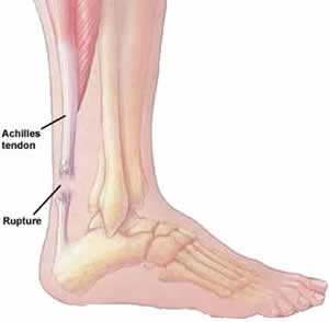
The Achilles tendon is the strongest tendon in the body but it is also the one that is most commonly injured. In this article we discuss the anatomy of the Achilles tendon as well as give an overview on Achilles tendon ruptures.Achilles Tendon Anatomy
The Achilles tendon is approximately 15 cm in length and is made up of a combination of fibers that are derived from two calf muscles which twist 90o and insert at the back of the calcaneus or heel bone. The Achilles tendon gets its blood supply from 3 sources. There is an area on the Achilles tendon approximately 2 to 6 cm above its insertion to the calcaneus that has poor circulation and is called the watershed region. Due to the diminished blood supply, this is a common area for Achilles tendon lesions and ruptures.
Acute Achilles tendon ruptures
Achilles tendon ruptures occur most often in middle-aged men during athletic activities. Patients with Achilles tendon ruptures often say that they felt a sudden pop or snap in their calf with subsequent weakness or difficulty with ambulation. Some say they felt someone kicked them or hit them from behind but there is noone in the vicinity. Patients may be able to walk with minor swelling and pain after a tear but have weakness.
Achilles tendon ruptures occur most often in middle-aged men during athletic activities. Patients with Achilles tendon ruptures often say that they felt a sudden pop or snap in their calf with subsequent weakness or difficulty with ambulation. Some say they felt someone kicked them or hit them from behind but there is noone in the vicinity. Patients may be able to walk with minor swelling and pain after a tear but have weakness.
Achilles Tendon Tear Exam
On examination, a palpable defect in the tendon can be felt and there is often an increase in upward movement of the ankle due to the lack of tension from the Achilles tendon. A common test that is used to evaluate the integrity of the Achilles tendon is the Thompson calf-squeeze test. Squeezing the calf muscles on the affected side will yield little to no movement in the ankle when compared to the unaffected side.
On examination, a palpable defect in the tendon can be felt and there is often an increase in upward movement of the ankle due to the lack of tension from the Achilles tendon. A common test that is used to evaluate the integrity of the Achilles tendon is the Thompson calf-squeeze test. Squeezing the calf muscles on the affected side will yield little to no movement in the ankle when compared to the unaffected side.
Imaging of Achilles Tendon Tears or Ruptures
MRI and ultrasound are often used to confirm the presence of an Achilles rupture and to evaluate the extent of the rupture. The diagnosis of Achilles tendon ruptures, however, is based on clinical examination. Imaging is often used for surgical planning or in cases in which there are equivocal examination findings.
MRI and ultrasound are often used to confirm the presence of an Achilles rupture and to evaluate the extent of the rupture. The diagnosis of Achilles tendon ruptures, however, is based on clinical examination. Imaging is often used for surgical planning or in cases in which there are equivocal examination findings.
Achilles Tendon Tear Surgical Advancements
Operative and nonoperative methods offered by foot and ankle doctors have shown favorable outcomes. Generally with surgical repair, there have been reports that there is a decreased rate of re-rupturing the tendon, improved strength, improved ankle motion, better return to activities, and fewer complaints.
Operative treatment involves reapproximating the two ends of the ruptured tendon together using special suture techniques. A repair can be augmented with a tendon transfer, or synthetic graft. Rarely, the Achilles tendon may rupture at its insertion into the calcaneus. In these cases, a tendon anchor may be used to reattach the Achilles to the bone. The patient is then put in a non-weightbearing cast for 4 to 6 weeks followed by gradual weight bearing and physical therapy.
No comments:
Post a Comment