Tuesday, March 26, 2013

Runner's Knee


A new course record was set at the Chicago Marathon last year.  With a time of 2:04:38, Tsegaye Kebede became the first Ethiopian man to win on Chicago’s flat course.  Thousands of first-timers and other runners followed the elite runners this morning.  For these endurance athletes, training is essential to reach the finish line, but overtraining or overpacing can easily lead to injury.  One common running injury is known as runner’s knee (patellofemoral pain syndrome) which is associated with pain behind the kneecap.  It is an overuse injury often seen in runners and cyclists, but it can also be seen in sports which require repetitive jumping or cutting.  While this pain can be cause by a direct impact injury, it is typically caused by repetitive bending of the knee.
 
The major symptom is pain which usually begins as a dull ache or stiffness behind the kneecap (patella).  The patella is attached to the thigh by the quadriceps muscle and tendon and to the leg by the patellar tendon.  Injuries or weakness of these supporting structures can lead to improper alignment and tracking of the patella as the knee  bends and straightens.  This results in irritation and pain.
 
Conservative treatments for patellofemoral pain syndrome begin with reduced training, ice, and anti-inflammatory medication.  Incorporate stretching and strength training of the quadriceps and hamstrings to improve stability around the knee.  In addition to these therapies, custom arch supports (orthotics) may be considered.
 
Recent studies have provided evidence that orthotics reduce patellofemoral pain by improving the tracking of the patella during the bending motion of the knee.  Although orthotics have been generally thought to benefit individuals with excessive collapsing of the arch (over pronation), these studies have shown that people of all foot types have reduction in knee pain with orthotics when compared to flat inserts.
 
Patellofemoral pain syndrome is a degenerative condition that can progressively get worse over time.  Proper training and conditioning can help prevent this and other lower extremity injuries.  Following any injury, the goal of returning to activity as quickly and safely as possible can be achieved with proper evaluation and treatment.  Custom orthotics casted by a podiatrist can be an important addition to the standard treatments for runner's knee.

Monday, March 25, 2013

Big Toe Joint Aches and Pains - What you need to know:


Often times patients will present with what they consider to be a "Bunion" in their big toe joint, and they attribute this to their pain. Many times, this is false. A bunion is an actual deviation or dislocation of the big toe joint, whereas the painful prominence which are often gradually increasing in size after many years removed from an injury can be related or directly attributed to progressive "bump" production which has the general buzzword "Arthritis".

Arthritis is actually a misnomer, because many times the inflammation of the joint is only noted initially in the disease process and over time the cartilage wears away forming spurs and bone prominence which leads to the pain and inability to move the joint fluidly. This is actually an "arthrosis" which is the end result of the condition. Arthritis is not usually visualized directly on a radiograph but arthrosis is easily visualized.

A clinician will find effusions whenever arthritis is active. This is the "Swelling" that patients often note is painful and warrants medical attention. This is often including joint space widening in the case of inflammatory arthritis but narrowing over time with osteoarthritis, and reduction of ability to move the joint without pain and with eventual grinding.

Patients will often times not recall the traumatic event which sparks the progressive wear down of this area on their foot, but many times this is not necessary. Patients will also note that the "bunion" is growing and is usually located on top and side of the joint. This is not a bunion, as these tend to only be located on the side of the joint and not at all on top.

What do you need to know to determine what to do next??

1) If the pain is at the end of the day you should see a foot and ankle specialist.

2) If the motion of the joint is limited or not similar to your other foot, you need to see the specialist.

3) If you have been given 2 or more cortisone injections in the same joint, you need to see a specialist.


There is a common theme here. A foot and ankle specialist is paramount to help a patient achieve a good result in treating first toe joint arthrosis. Often times, this is progressive and may limit treatment options depending on the stage of the pathology. There is no actual cure for any form of arthritis, but there are means to treat and accommodate the condition in order to reduce pain and limitation with walking. We are adept in treating all forms of arthritis, from the initial swelling stage, to the end stage limited motion in the toe joint. There are many times several good options depending on the stage of the disease process, and we will be able to give you a definitive solution in many cases.

Advancement in Achilles Tendon Tear and Achilles Rupture Care

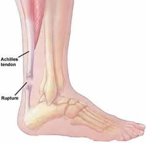
The Achilles tendon is the strongest tendon in the body but it is also the one that is most commonly injured. In this article we discuss the anatomy of the Achilles tendon as well as give an overview on Achilles tendon ruptures.
 
Achilles Tendon Anatomy

The Achilles tendon is approximately 15 cm in length and is made up of a combination of fibers that are derived from two calf muscles which twist 90o and insert at the back of the calcaneus or heel bone. The Achilles tendon gets its blood supply from 3 sources. There is an area on the Achilles tendon approximately 2 to 6 cm above its insertion to the calcaneus that has poor circulation and is called the watershed region. Due to the diminished blood supply, this is a common area for Achilles tendon lesions and ruptures.


Acute Achilles tendon ruptures

Achilles tendon ruptures occur most often in middle-aged men during athletic activities. Patients with Achilles tendon ruptures often say that they felt a sudden pop or snap in their calf with subsequent weakness or difficulty with ambulation. Some say they felt someone kicked them or hit them from behind but there is noone in the vicinity. Patients may be able to walk with minor swelling and pain after a tear but have weakness.
Achilles Tendon Tear Exam

On examination, a palpable defect in the tendon can be felt and there is often an increase in upward movement of the ankle due to the lack of tension from the Achilles tendon. A common test that is used to evaluate the integrity of the Achilles tendon is the Thompson calf-squeeze test. Squeezing the calf muscles on the affected side will yield little to no movement in the ankle when compared to the unaffected side.
Imaging of Achilles Tendon Tears or Ruptures

MRI and ultrasound are often used to confirm the presence of an Achilles rupture and to evaluate the extent of the rupture. The diagnosis of Achilles tendon ruptures, however, is based on clinical examination. Imaging is often used for surgical planning or in cases in which there are equivocal examination findings. 
 
Achilles Tendon Tear Surgical Advancements

Operative and nonoperative methods offered by foot and ankle doctors have shown favorable outcomes. Generally with surgical repair, there have been reports that there is a decreased rate of re-rupturing the tendon, improved strength, improved ankle motion, better return to activities, and fewer complaints.

Operative treatment involves reapproximating the two ends of the ruptured tendon together using special suture techniques. A repair can be augmented with a tendon transfer, or synthetic graft. Rarely, the Achilles tendon may rupture at its insertion into the calcaneus. In these cases, a tendon anchor may be used to reattach the Achilles to the bone. The patient is then put in a non-weightbearing cast for 4 to 6 weeks followed by gradual weight bearing and physical therapy. 

Orthotics


Finally when the perfect pair shoes are chosen, they like the style, size, and choice of materials, the important stuff can happen, getting your custom orthotics from your local podiatrist. Orthotics is a custom foot support, which you place in your shoes for your pain free style. Custom orthotics are designed specifically for you, it gives a better arch support then the regular over the counter (OTC) kind. It does this by correcting the biomechanics of your walking style, to overall help support your feet, knees and legs.

Custom orthotics are useful for people: who are overweight; obligated to be on their feet for extended periods of time; or have a previous foot or ankle issues. Even athletes use them to optimize performance and enhance comfort while either in training session or on game days. Patients with arthritis or diabetic foot conditions use them for comfort and protection. Orthotics helps prevent injury or hinder the progression of existing problems, thus allowing healing to take place. On the other hand, even if no major conditions or symptoms exist, orthotics provides protection and comfort for your feet.

If you or someone you know hasn’t had a chance to get their custom orthotics this year it is time to see your podiatrist and be evaluated and casted today. Whether it’s your first or third pair I would be more than happy to schedule an appointment. We will be able access your walking style, and design and create a perfect custom orthotic for your pain free style.